Dosimetry Protocols for Lu-177 Therapies
Comprehensive guidelines for Lu177 dosimetry and calculations.
General SPECT/CT Setup
Gamma Camera: Siemens Symbia SPECT/CT
Collimator: MELP
Energy Window Settings
SPECT Acquisition Parameters
Projections: 60 (120 images)
Angular Resolution: 3°
Acquisition Time: 10 seconds per projection (Extendable to 20 sec for late time points)
Matrix: 128 × 128
Zoom: 1.0
Contour Mode: Auto-contour
Motion: Continuous or Step & Shoot
CT Acquisition
Tube Voltage: 120–130 keV
Tube Current: 30–40 mAs
CTDIvol approx. 3.0
Image Reconstruction
Method: Hybrid Reconstruction
Iterations/Subsets: 5 iterations, 16 subsets
Post-Filter: None
Corrections Applied:
Resolution Recovery (RR)
Attenuation Correction (AC)
Scatter Correction (SC)
Calibration Factor: 9.8
Imaging Time Points and Dosimetry Tools
1.PSMA targeted therapy
2.DOTATATE therapy
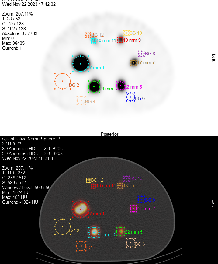
Quantitative Tomographic NEMA Sphere Phantom Validation Measurement
Aim
To perform a SPECT validation using a NEMA sphere phantom.
Instrumentation
IEC NEMA Body phantom with 6 spheres
Additional sphere with at least 6 cm in diameter
500 mL beaker
Flat head screwdriver
Funnel
Filling needle
~400 MBq Lu-177
~10 L of pure water
0.2 mmol citric acid
Methods
Fill Phantom with Water
Attach the spheres and fill the phantom with pure water.
Do not place a lung insert in the phantom.
Insert the lid with spheres attached into the phantom.
Position the largest sphere at the 3 o’clock position, with decreasing sphere sizes in clockwise direction when viewed from the top.
Prepare Phantom with Lu-177
Measure the patient vial in the dose calibrator and note the activity value.
Handle Lu-177 under a fume hood. Leave the vial under an extraction fan for 15 minutes.
Fill approximately 100 mL of the solution into the beaker.
Add around 0.2 mmol citric acid to prevent sticking.
Remove the screws from each sphere in preparation for filling.
Draw ~400 MBq of Lu-177 into a syringe and measure the activity in the dose calibrator.
Record Syringe Activity Pre-Filling
Source: Syringe pre
Volume [ml]:
Activity [MBq]:
Date:
Time:
Add the Lu-177 activity from the syringe to the beaker. Flush several times to recover all activity using the same solution.
Refill the syringe with saline or water to initial volume and measure residual activity.
Record Syringe Activity Post-Filling
Source: Syringe post
Volume [ml]:
Activity [MBq]:
Date:
Time:
Measure the remaining activity in the vial.
Record Vial Residual Activity
Source: Patient Vial post
Volume [ml]:
Activity [MBq]:
Date:
Time:
Top up the stock solution to 200 mL using balance or measuring cylinder.
Stir the beaker solution for 30 seconds.
Draw the solution into a clean syringe and fill each sphere, avoiding air bubbles.
Seal the spheres after filling.
Fill the 6 cm sphere from the same stock solution (either now or later, as applicable).
Stock Solution Parameters
Volume in stock solution:
Volume determined by:
Balance
Measuring Cylinder
Perform SPECT/CT Scan
Position camera heads in H-mode with the phantom centered in the field of view.
Save the scanner's energy spectrum and attach it to the worksheet.
Acquire a scan using the protocol "CAAA617B12302_SPECT".
Scan Target
The scan should acquire 3 million counts in the 208 keV window.
Scan Parameters
Study start date:
Study start time:
Total counts acquired for 208 keV: 3 million
Total scan duration:
Notes:
Perform local reconstruction using OSEM iterative algorithm.
Apply CT-based attenuation and scatter correction.
Use the same reconstruction algorithm intended for patient imaging.