Recovery Coefficient Analysis Using NEMA Phantom
📌 Introduction
Accurate quantification of radiopharmaceutical uptake in small lesions is critical for effective radionuclide therapy planning. However, due to limited spatial resolution in SPECT/CT imaging, Partial Volume Effects (PVE) lead to underestimation of activity concentration. This study evaluates the Recovery Coefficient (RC) under different Sphere-to-Background Ratios (SBR) using a NEMA IEC Body Phantom with Lu-177.
🧪 Methodology
Phantom Setup
Phantom Type: NEMA IEC Body Phantom with 6 fillable spheres
Tracer: Lu-177 (~400 MBq)
Sphere Volumes: 0.52 ml to 26.52 ml
Medium: Distilled water + citric acid to avoid wall binding
Acquisition: Siemens SPECT/CT using the 208 keV window
Reconstruction: OSEM with attenuation and scatter correction
SBR Conditions
The same sphere concentration was maintained across all setups:
Sphere Concentration: 3.85688 MBq/ml
Background concentrations were adjusted to create different SBR levels:
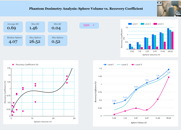
📊 Results
Recovery Coefficient by Sphere Volume and SBR
The Recovery Coefficient (RC) was calculated as:
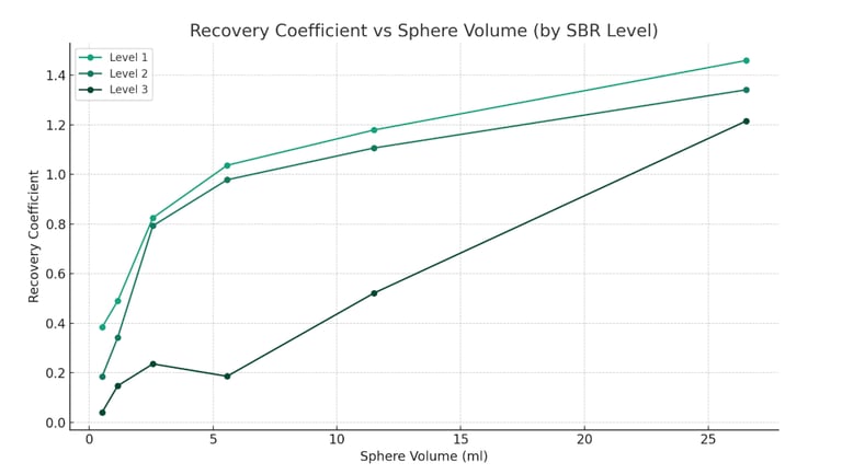
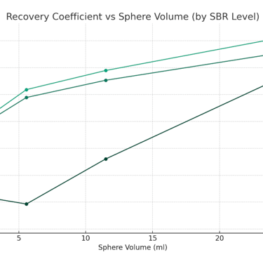
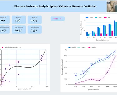
Recovery Coefficient vs. Sphere Volume for the three different background levels (SBR levels):
Level 1 (high contrast) shows the highest RCs, especially for small spheres.
Level 2 has moderate RC values, showing more partial volume loss.
Level 3 (low contrast) demonstrates the strongest partial volume effects, with low RCs in small spheres.
This graph clearly visualizes how partial volume effects increase as contrast (SBR) decreases, especially in small lesions.